
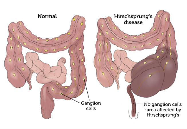
Hirschsprung's Disease (HH) is a disease characterized by the absence of parasympathetic ganglion cells in the myenteric and submucosal nerve plexuses of the colon, and the inability to relax in the affected bowel. It usually affects the rectosigmoid region, which is the last part of the large intestine. Very rarely, it can involve the entire large intestine and part of the small intestine. In the affected part, the intestine cannot relax and remains contracted and this area is blocked. As a result of the obstruction, the stool cannot progress and the healthy intestine works hard to advance the stool, which causes the healthy intestine to expand and thicken. The result is a narrow distal segment and an enlarged and thickened proximal segment. Hirschsprung's Disease occurs in one in 5000 births and is more common in boys. The exact cause of Hirschsprung's Disease is not clear.
Symptoms of Hirschsprung's Disease
Constipation and abdominal distention are the most common findings, although they vary in the newborn and pediatric age group. The longer the segment involved, the greater the severity of the complaints and the disease. Meconium output may not be observed in the first 24 hours of life in 90% of newborns with HH. On physical examination, fecal discharge with rectal touch is typical.
Diagnosis in Hirschsprung's Disease
One of the important methods in diagnosis is colon radiography. During this procedure, the intestine is visualized by administering diluted barium from the anus to the large intestine. In Hirschsprung's disease, the bowel is narrow in the contracted aganglionic part. When the drug is continued to be given, the slowly enlarged part of the intestine appears. The presence of narrow and large intestinal segments is descriptive of Hirschsprung's disease.
The main diagnostic method in Hirschsprung's disease is full-thickness rectal biopsy. For this, a full-thickness biopsy is taken from the rectum under operating room conditions. The biopsy sample taken is examined under the microscope with special staining methods and it is checked whether there are ganglion cells. In Hirschsprung's disease, there are no cells between the muscle tissues and the nerve structures found here are larger than usual (hypertrophic nerve plexuses).
Suction biopsy: It is less invasive than full-thickness rectal biopsy, but its accuracy rate is lower. It is preferred in many centers.
Another method used for diagnosis is the manometric study. In this study, the contraction and relaxation of the muscles in the last part of the large intestine and around the anus, depending on the stimulus, are investigated with special pressure catheters placed on the anus and just above it. It is diagnostic that there is a natural continuous contraction in the narrow aganglionic segment and that this area does not relax.
Treatment in Hirschsprung's Disease
There is no cure for Hirschsprung's disease other than surgery.
In the treatment, the aganglionic cut is removed in the surgery and replaced with the ganglionic cut above it and connected to the anus or the intestine just above it (pull-through process).
surgical procedure; It is performed in one, two or three stages, depending on the age of the child, the length of the aganglionic segment and the findings.
Three-phase method: It is a method that was first defined and still used today. Accordingly, in the first stage, the part of the large intestine with ganglion cells is temporarily attached to the abdominal wall. With this method, called colostomy, it is aimed to rest the enlarged intestinal segment and allow the child to easily remove the stool. In the second phase; The gut without ganglion cells is removed or disabled and replaced with the gut with ganglion cells. In the third stage, the colostomy is closed and the treatment is completed.
Two-stage surgeries: In the first stage, the part of the large intestine with ganglion cells is temporarily attached to the abdominal wall. In the second phase; The intestine without ganglion cells is removed or disabled and the colostomy tip with ganglion cells is released from the abdominal wall and pulled down towards the anus.
Single-stage surgeries: It can be performed as an open surgical method or closed (laparoscopically). In both methods, the intestinal tissue with ganglion cells is identified by a rapid pathological examination (frozen) during the operation, and the operation is completed by pulling this area down to the anus.
Primary Transanal Pull_Through (Primary TEP): It is a new method that has been widely used in recent years. It is in the form of entering directly through the anus without opening the abdomen of the patient, pulling the aganglionic intestine out from here and connecting the ganglionic intestine to the anus.


